Bone protection for DMD patients taking corticosteroids

Corticosteroid treatment increases the risk of vertebral fractures in all children including those with DMD, although the precise risk for this population is not known. Vertebral fractures can cause no symptoms and because they are not routinely screened for, they are not recognised, thus only those fractures causing pain are detected. The definition of a vertebral fracture is not straightforward, at the mildest end of the spectrum there may be only a slight loss of height of the vertebrae while at the severest end the vertebra appears wedged and narrow in shape. Fractures of the vertebrae either occur spontaneously or following a fall onto the buttocks.

In healthy children there is an increase in bone mineral strength with advancing age, corticosteroid treatment interferes with this process. In children with DMD this effect may be less pronounced with intermittent than daily treatment. However, when walking is lost, there is a sharp decrease in bone strength and the risk of vertebral fractures is increased for all steroid regimens. Switching from an intermittent to daily steroid regimen at the point at which walking becomes more difficult may accelerate the risk of vertebral fracture.

Vertebral morphometry is a computerised method of measuring vertebral shape and height from an X ray of the spine and is one way in which subtle changes in vertebral shape can be monitored. The other way to study bone health is by a DEXA scan which measures Bone Mineral Density (BMD). The relationship between these techniques and fracture risk in DMD is not known, although these data are emerging for children as a whole.

Puberty is a time when bone strength normally increases due to hormones such as testosterone. Delayed puberty is an additional problem for steroid treated children, thus if the child is not showing signs of puberty by the age of 14 years then referral to a paediatric endocrinologist for testosterone treatment should be considered.

Weight bearing is important for maintaining bone strength, which is why fracture risk increases dramatically when walking stops, thus, maintaining walking or standing is important in children with DMD. Vitamin D helps to maintain strong bones, adequate blood levels of vitamin D should be maintained for all children taking steroids by giving a daily vitamin D supplement. Calcium is also important for bone health and has a better effect when taken as dairy products, unless the child has a poor dairy intake, in which case a supplement should be given. Bisphosphonates are a group of drugs that can improve bone strength and reduce pain following a fracture. Currently, they are used in children to treat fractures but they are not routinely used to prevent fractures. This is because bisphosphonates remain in bone tissue for many years and the long term effects are not known, although this data may be available soon from studies in children with other conditions.

Bone markers are blood and urine tests which can give an idea of the degree of bone loss or laying down of new bone. Their relationship with the risk of fracture is not known, but they can be useful to monitor the effects of Bisphosphonate treatment. The workshop consensus was that a DEXA scan of the lumbar spine should be performed annually in all DMD children receiving steroids and if the bone mineral density Z score ( adjusted for body size) is less than or equal to -2.0, an X ray of the spine should be performed to look for vertebral changes. Any changes of vertebral shape should be discussed with a metabolic bone expert for consideration of preventative treatment with a Bisphosphonate.

Children presenting with spontaneous painful vertebral fractures should be treated with a bisphosphonate. On the other hand, children who have a single painful vertebral fracture following a fall on the buttock will require pain relief but may not necessarily require treatment with a bisphosphonate.

A full report is published in Neuromuscular Disorders (pdf)
Rare Structural Congenital Myopathies

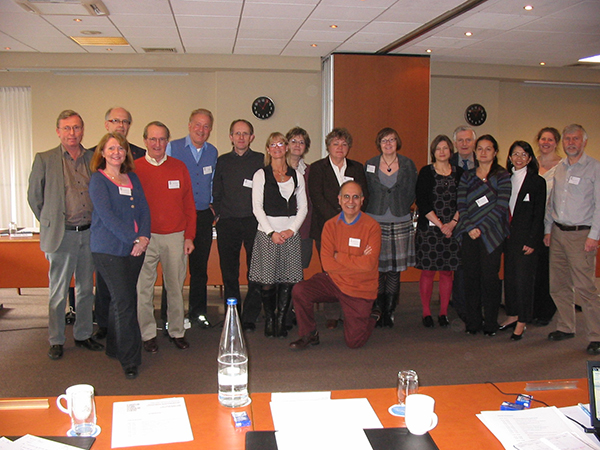
Seventeen clinicians and scientists from six European countries, Argentina, Japan, and the USA recently assembled for the 169thENMC-sponsored International Workshop on “Rare Structural Congenital Myopathies” in Naarden, The Netherlands. The aim of the workshop was to identify sufficient informative families and muscle tissue specimens in this group of neuromuscular conditions to analyse their genetic causation either by linkage analysis (a technique to trace a genetic condition within families) in informative families or by morphological studies on muscle tissues, including laser capture microdissection (a microscopic technique to directly isolate the abnormal structures from affected muscle) analysis to lay the foundation for future therapeutic approaches.

Background:

Among the congenital myopathies, there are more frequent and classical ones, such as nemaline myopathies, core diseases, and centronuclear myopathies for which dedicated workshops have already taken place at ENMC and consortia are in existence. However, there still are other, so called “rare structured congenital myopathies”, which so far had only been addressed once before, at the 56th ENMC International Workshop in 1997. With the tremendous progress in new gene identification in the congenital myopathies and with the development of novel microscopic, genetic and biochemical technologies and with the establishment of dedicated European and worldwide research networks, such “rare structured congential myopathies” have now come into focus again. Amongst those myopathies, the 169th ENMC International Workshop focused on tubular aggregate myopathy, cylindrical spirals myopathy, Zebra body myopathy, fingerprint body myopathy, and crystalline body myopathy.

Workshop contents:

Because these congenital myopathies are very rare individually, the recruitment of a informative families and/or muscle tissue specimens is critical to proceed towards their molecular analysis. The workshop achieved this goal by assembling clinicians and scientists from several major international neuromuscular institutions, not only from Europe but also from the USA, South America, and Japan who had reviewed their extensive archives to locate cases, informative families, and muscle tissue specimens.

Conclusion:

Having reviewed worldwide data on these rare myopathies, a consortium has been formed to move forward towards a comprehensive analysis of patients, families and muscle tissue, applying specialized techniques such as the use of antibodies (immunohistochemistry), laser capture microdissection analysis, and advanced gene analysis technology.

Only when genes and mutations will have been identified in these afore-mentioned “rare structural congenital myopathies”, a meaningful therapeutic approach can be conceived and hopefully brought back to the patients.

A detailed report on this workshop is published in Neuromuscular Disorders (pdf).

Participants:

Boersen, A. (ENMC)

Bönnemann C.G. (Philadelphia/USA)

Bertini, E. (Rome/Italy)

Claeys, K. (Paris/France)

Figarella-Branger, D. (Marseille/France)

Goebel, H.H. (Mainz/Germany)

Holton, J. (London/UK)

Hübner, A. (Dresden/Germany)

Kirschner, J. (Freiburg/Germany)

Lammens, M. (Nijmegen/The Netherlands)

Moore, S. (Iowa City/USA)

Nonaka, I. (Tokyo/Japan)

Olivé, M. (Barcelona/Spain)

Pegoraro, E. (Padova/Italy)

Polvikoski, T. (Newcastle/UK)

Schessl, J. (Munich/Germany)

Sewry, C. (London/U.K.)

Taratuto, A. (Buenos Aires/Argentina)

Clarke, N. (Sydney/Australia) submitted data to be presented at the workshop.

Outcome Measures and Clinical trials in CHARCOT-MARIE-TOOTH DISEASE

The 168th ENMC workshop organized from 18 – 20 September 2009 in The Netherlands was the second ENMC workshop on Charcot-Marie-Tooth. The first one was the 136th ENMC workshop held in April 2005.

Twenty-three participants from 9 countries (Australia; United Kingdom; France; Germany; Italy; Spain,Belgium, U.S.A. and The Netherlands) attended this ENMC workshop on Outcome Measures and Clinical Trials in Charcot-Marie-Tooth disease. Participants included adult and paediatric neurologists, biologists, molecular geneticists, two allied health professionals and representatives from TREAT NMD and two CMT patient organisations (Italian and US).

The ENMC workshop on outcome measures and clinical trials in CMT was very successful.

The main aims of the workshop were:

1) to discuss and agree how to modify the CMTNS for adults to increase sensitivity following the experience with the Vitamin C trials.

2) to prepare a draft paediatric impairment and disability scale for children with CMT.

3) to discuss an international CMT patient registry done in collaboration with Treat-NMD and to agree a minimal dataset

4) to discuss trial design for CMT.

Achievements.

1) During the workshop we worked on a modified version of the CMTNS for adults and plan to finalise and validate this version.

2a) A draft paediatric score was prepared and this will be further modified and validated over the next year.

2b) Furthermore, a joint ENMC working group was established to develop and test CMT-specific QoL measures for children and adults.

3) The workshop was enthusiastic that an international CMT patient registry be developed in collaboration with Treat-NMD and a minimal dataset is being developed. Furthermore, the facility is available to host the registry in the University of South Florida, USA.

4) A very useful discussion on trial design for CMT took place based on our experience with Vitamin C trials and the importance of powering trials sufficiently, choosing appropriate primary outcome measures, running trials for an adequate length of time, proper preclinical and early clinical trials, and biological markers evaluation was emphasized. A full report is published in Neuromuscular Disorders(pdf)

PENTACON DIGITAL CAMERA

X-linked myopathy with excessive autophagy (XMEA)

The 167th ENMC workshop organized June 19-21,2009 in Naarden, The Netherlands was the second (= follow-up) workshop on X-linked myopathy with excessive autophagy (XMEA) and related vacuolar myopathies, the first one having been the 77th ENMC workshop March 10-12, 2000. The initiative to organize this follow-up workshop was the fact that the gene defects which cause XMEA and the main features how the myofibers are damaged (Ramachandran, Munteanu et al, Cell 2009; 137:235-246) have just recently been clarified. The workshop comprised of 16 clinicians/clinical researchers and basic scientists from 4 ENMC member states and 4 non-member states.

Vacuolar myopathies comprise a group of several different muscle diseases, both hereditary and acquired. They have in common the presence of vacuoles within the sarcoplasm of the diseased myofibers. It has become evident that the structure of the vacuoles is not the same in different vacuolar myopathies and their structure reflects the reason why vacuoles are formed. XMEA belongs to such a group of vacuolar myopathies in which the vacuoles have been defined as autophagic vacuoles with sarcolemmal features (AVSF). This means that the vacuoles are lined by a membrane that is similar as the sarcolemma, i.e. the membrane that lines the whole muscle fiber. Autophagy may be called the recycling system of the cells: In short, molecules and cellular organelles, which have faulty structure or are worn out, are isolated into membrane bound vacuoles, so-called autophagic vacuoles. Within another membrane bound vacuole called lysosome the cell transports into autophagic vacuoles enzymes, which are able to break down (degrade) the “trash” (= cell debris) within the autophagic vacuoles. Many of the breakdown products are then re-used by the cell. An important feature is the fact that the degradative enzymes require acid surroundings (low pH) to function and therefore hydrogen ions must be pumped into the autophagic vacuoles to lower the pH to a correct level.

So far the genetic defect has been discovered in two AVSFs: XMEA is one of them and Danon´s disease with mutation in LAMP-2 (a protein in the membrane of lysosomes) is the other. The gene defect in XMEA is unique among myopathies. The defect is in VMA21 gene, the protein product of which is needed in the construction of a protein complex V-ATPase, which is responsible for pumping hydrogen ions into lysosomes, i.e. making the pH within lysosomes acidic. In XMEA the defect inVMA21 results in too little amount of V-ATPase, consequently too few hydrogen ions are pumped into the autophagic vacuoles (= too high pH) and therefore the cell debris within the autophagic vacuoles cannot be degraded but instead it accumulates within the myofibers.

At present 13 unrelated families have been diagnosed to have XMEA with VMA21 mutation. In this workshop the clinical pictures of XMEA patients in different countries were compared. It was in principle similar, though the age of onset and rate of progression varied to some extent. Most often it begins in childhood and is relatively slowly progressive, though even congenital cases have been diagnosed in Japan. The EMG finding of myotonic discharges without clinical myotonia could be detected even in clinically asymptomatic patients. The MRI findings appear to be quite suggestive of XMEA. The muscle biopsy findings are very uniform and multilayered basal lamina as well as extrusion of the debris out of the myofibers (exocytosis) appear to be unique features of XMEA, which differentiates it from other AVSFs, some with identified gene defects like Danon disease, some with known linkage to chromosome 19 and some with no genetic data available so far.

An obvious therapeutic possibility in XMEA would be the normalization (lowering) of the pH within the autophagic vacuoles. This possibility was discussed at the workshop, but since the gene defect and the pathogenesis of XMA have been published only this April, therapeutic experiments are just being planned.

This workshop was organized by Prof. H. Kalimo and held in Naarden in June 2009.
The report for this workshop is not (yet) available. For further information, please contact one of the workshop organizers or the ENMC office.

Collagen VI related myopathies

Twenty-four participants from 8 countries (Australia; England; France; Germany; Italy; Switzerland; Turkey; USA) attended an ENMC workshop on the topic of collagen VI-related myopathies which include the diseases known as Ullrich congenital muscular dystrophy (UCMD) and Bethlem Myopathy (BM). Participants included biochemists, biologists, molecular geneticists, neurologists, paediatricians and representatives of a patient advocacy organization and pharmaceutical companies.

Collagen VI-related myopathies are a group of neuromuscular disorders due to mutations in any of the three genes encoding collagen VI (ColVI), COL6A1COL6A2 and COL6A3. Defects in ColVI, a major extracellular matrix component in many tissues, were initially reported in BM and UCMD which were considered as two separate entities with different patterns of inheritance. In the last few years, the clinical picture has greatly expanded. The results of studies in models of the disease indicate an opportunity for therapeutic interventions, one of which has recently been evaluated in a pilot trial. Two larger clinical trials are under consideration in partnership with industrial partners.

The present meeting covered areas from genetics and the clinical phenotypes associated with ColVI defects, to the molecular processes taking place in damaged tissue and the therapeutic avenues that will be investigated in the upcoming clinical trials. Several participants reported on the complexity of the molecular diagnosis and the emergence of novel approaches to detect complex gene rearrangements. The participants have recognized the need for a revised clinical classification taking into account patients with intermediate presentation. In addition there are clearly some patients with phenotypes highly suggestive of UCMD or BM where no ColVI mutations can be found, so other genes are likely to be involved. The need to study other candidate genes was discussed, in particular the recently describedCOL6A5 and COL6A6 genes whose expression has been refined in various tissues.

Studies on the mechanisms linking ColVI mutations to muscle pathology were reported in models of the disease and in cell cultures derived from patients, and the possibility for therapeutic interventions was discussed, along with the need for relevant outcome measures to be taken into consideration for clinical trials.

Currently existing patient registries and plans for an international collaborative effort with the patient organization Cure-CMD and the TREAT-NMD network were presented.

A full report is published in Neuromuscular Disorders (pdf)
Distal Myopathies

The ENMC consortium on distal myopathies held its 3rd workshop in Naarden, the Netherlands, February 6-8th, 2009. It was attended by 23 active participants from Finland, Japan, Israel, France, Germany, Australia, Sweden, Switzerland, Italy, Spain, Austria, UK and the USA. During the last 15 years the field of known and defined distal myopathies has expanded from a handful clinical entities to more than 16 genetically defined different diseases. For the majority of these diseases the underlying gene defect is established and the further disease mechanisms on the cellular level are currently explored. Due to this remarkable expansion in the number of distal myopathies the 3rd workshop was concentrating on the diseases with involvement of the feature of the so called rimmed vacuoles on muscle biopsy and/or diseases caused by defects in genes needed for proteins in the contractile machinery of the muscle cell, the sarcomeric proteins.

The participants reported on a number of new mutations and new variations in the clinical features of the known distal myopathies. Other reports detailed newly discovered genes underlying distinct forms of distal myopathy, some of them yet unpublished. Considerable progress has been made in the clarification of the defects on the protein level and the molecular processes taking place in the muscle cells damaged by the mutations, even though these processes are very complex and not yet fully understood. Based on the existing knowledge some treatment options have already been tested in disease models for a few forms of distal myopathy, such as the GNE-mutated HIBM/DMRV disease. The results of these cellular and animal studies indicate a therapeutic opportunity that may be applicable to the human disease and soon open the way for clinical trials in patients. The workshop agreed on guidelines for diagnostic procedures for the clinicians, emphasizing the role of muscle imaging to complement and direct the diagnostics towards a molecular genetic diagnosis. These guidelines will be included in the extended report of the meeting to be submitted for publication in Neuromuscular Disorders.

A detailed report of these conclusions is now published in Neuromuscular Diseases (pdf)

Centro Nuclear Myopathy

18 participants from different countries convened for the 164th ENMC International workshop on myotubular/centronuclear Myopathies, a group of inherited muscle disorders with a wide spectrum of severity and associated features. Although to date changes in a number of different genes have been identified to cause myotubular/centronuclear myopathy, there is a substantial proportion of patients with the condition where the underlying cause remains currently unknown. The first aim of this workshop was to delineate and further clarify the features associated with forms of the disorder where the underlying gene defect is already known, and to outline common features in forms of myotubular/centronuclear myopathy without known genetic defect in order to identify patients for further study. The clinical part of the workshop was complemented by sessions on the biochemical effects of specific genetic changes and on animals suffering from similar conditions; those may present suitable models to further understanding of myotubular/centronuclear myopathy and to study potential treatments. Another session focused on the design and establishment of databases collecting information from patients suffering from myotubular/centronuclear myopathy, a prerequisite for collecting patient populations for future therapy trials and a beneficial tool for distributing information about these rare conditions to those affected by them and their treating clinicians. During the final session of the workshop potential therapeutic approaches to myotubular/centronuclear myopathy based on findings in an animal model of the condition were presented and discussed. Recommendations for a structured approach to the genetic diagnosis of the condition and on the establishment of a patient registry for myotubular/centronuclear myopathy were made and strategies to implement those were discussed. Lastly, plans for further collaborations and meetings were drafted.

A full report is published in Neuromuscular Diseases (pdf)