Developing guidelines for management of reproductive options for families with maternally inherited mtDNA disease

Location: Hoofddorp, the Netherlands

Title: Developing guidelines for management of reproductive options for families with maternally inherited mtDNA disease

Date: 22 – 24 March 2019

Organisers: Dr J. Burgstaller (Austria), Dr. R McFarland (UK), Prof. J. Poulton (UK), Prof. J. Steffann (France)

Translations of this report:
Dutch by Dr S. Sallevelt
French by Prof. J. Steffann
German by Dr B. Arbeithuber
Spanish by Dr J. Bengoa

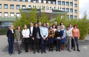
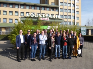
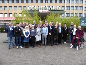
Participants: Dr. B. Arbeithuber (U.S.A), Dr. J. Bengoa (France), Dr. J. Burgstaller (Austria), Dr. S. Chan (UK), Dr. M. Chiaratti (Brazil), Dr. M. Crouch (UK), Dr. R. Dimond (UK), Dr. T. Enriques (Spain), Dr. G. Gorman (UK), Dr. L. Hyslop (UK), Dr. I.  Johnston (UK), Mr. and Mrs. J. Kitto (UK), Mrs. A. Maguire (UK), Dr. R. McFarland (UK), Dr. S. Mitalipov (USA), Mrs. Van Otterloo, (The Netherlands), Prof. J. Poulton (UK), Dr. S. Sallevelt (The Netherlands), Dr. H. Smeets (The Netherlands), Dr. C. Spits (Belgium), Prof. J. St. John (Australia), Prof. J. Steffann (France), Dr. J. Stewart (Germany), Dr. M. Stoneking (Germany), Prof. D. Thorburn (Australia), Ms. E. van der Veer (The Netherlands) and Dr. D. Wells (UK)

Background

Genetic counselling is uniquely difficult in the group of muscle diseases caused by shortage of energy  known as “mitochondrial diseases”. Mitochondria are small parts of cells that make energy.  They depend on their own unique blueprint, the mitochondrial DNA (mtDNA, the genetic material coding for the energy factories in each cell). MtDNA is inherited exclusively from the mother, and usually all of the mtDNAs in a normal person are identical.  In mitochondrial diseases patients may harbour both normal and damaged mtDNA (a situation called heteroplasmy) so babies may inherit both normal and damaged mtDNA from their mothers. Heteroplasmy makes it difficult to predict the chance that a mother with mitochondrial disease will transmit damaged mtDNA to her children. In addition, if the heteroplasmy level is measured in an unborn child it can be difficult to accurately predict how the child might be affected.

There are several different genetic options aiming to prevent the birth of a child affected by severe disease by reducing or preventing transmission of damaged mtDNA.

The simplest option is to offer a healthy egg donated by an unaffected woman.

A second option is pre-implantation genetic diagnosis (PGD).  In PGD a woman’s eggs are collected and fertilized with her partner’s sperm in a test tube.  A single cell is sampled from the embryos at an early stage in their development, and the healthiest embryo is placed in the woman’s womb.  PGD reduces but does not completely eliminate the risk of having an affected child, and cannot be offered to patients in whom all the mtDNA is mutated (homoplasmic).

While PGD is the established prevention procedure in most countries, mitochondrial replacement therapy (MRT) is available as a third option in the UK, and steps have been taken towards making it available in Australia. In the Netherlands MRT is legal but implementation is impractical because the current law prohibits generating embryos for research, which would be needed to set up the technique.

In MRT the nucleus is removed either from an embryo at the very early stages of development, or from an egg from the carrier female and placed into a healthy donor cell at the same stage from which the nucleus has been removed.

We aimed to develop guidelines for the new reproductive options that are becoming available for families with maternally inherited mtDNA disease.

What was achieved?

The scientific, social and ethical background leading to the introduction of MRT into genetic management were discussed.  The patients at the meeting felt that the debate had been generally useful. It revealed that in some regions, mitochondrial families had been given little information about the opportunities available.  The patient representatives who participated in the workshop are enthusiastic about MRT and feel that the potential benefits out-weigh the uncertainties of these new options.  A survey of adult patients in the UK who were consulted about the new technologies showed that patients were broadly supportive of legalization and this was the case even though the debates focused on severe childhood illness which did not match their own experience of late onset disease.  They also explained that some of the sound bites used by the UK press were unhelpful.

The need for continuing research was emphasized and participants from this workshop have made plans to collaborate in future projects. A full report of this ENMC workshop, including the scientific advances, will be published in Neuromuscular Disorders.

Outcomes: Counselling protocols and clinical guidelines of who can be offered MRT were discussed.

The following key deliverables were achieved:

  1. Consensus on referral of patients for MRT
  2. Counselling guidelines for units carrying out MRT

The clinical guidelines will be reported at international meetings including EUROMIT 2020.  A follow up ENMC meeting reporting on implementation of these guidelines in different countries and the short- and long term outcomes of the procedures is planned.  This information will be important for  countries discussing legalisation of these approaches.

In total, 29 people attended the ENMC workshop including participants from The Netherlands, UK, France, Germany, Spain, Austria, Belgium, Australia, USA and Brazil. The group was multi-disciplinary including patients, clinicians, basic scientists, ethicists, a sociologist, and representatives of industry and patient organizations (including the Lily Foundation, the Dutch Muscular Disease Association, International Mito Patients (IMP) and the LHON group of the Dutch Eye Association).

Glossary, suggestions for explanations can be found at https://www.musculardystrophyuk.org/about-muscle-wasting-conditions/glossary/ 
A full report is published in Neuromuscular Disorders (PDF).
 

Diagnosis and Management of Juvenile Myasthenia Gravis

Location: Hoofddorp, The Netherlands

Title: Diagnosis and Management of Juvenile Myasthenia Gravis

Date: 1-3 March 2019

Organisers: Dr P. Munot (UK), Dr E. Niks (The Netherlands), Dr J. Palace (UK) and Dr S. Robb (UK)

Translations of this report by:
Dutch by Dr E. Niks
German by Dr A. Della Marina

Participants: Dr F. Baggi (Italy), Dr M. Borresen (Denmark), Dr P. Cruz, Dr A. Della Marina (Germany), Dr C. Erasmus (The Netherlands), Prof. A. Evoli (Italy), Prof. B. Eymard (France),  Dr I. Hughes (UK), Prof. H. Jungbluth (UK), Dr A. Klein (Switzerland), Dr N. Kuntz (U.S.A.), Dr P. Munot (UK), Dr E. Niks (The Netherlands), Dr J. Palace (UK), Dr M. Pitt (UK), Dr S. Ramdas (UK), Dr S. Robb (UK) and Prof. M. Ryan (Australia)

The 242nd ENMC workshop was convened between 1 and 3 March 2019 and brought together a patient representative and neuromuscular experts from Europe, Australia and the United States to discuss the care of children with Juvenile Myasthenia Gravis. The aim was to develop guidance on diagnosis and management of Juvenile Myasthenia Gravis, after reviewing the latest evidence and collating expert opinion.

Juvenile myasthenia gravis (JMG) with onset of symptoms below the age of 18y, like its adult counterpart (MG), is a disorder of neuromuscular transmission, resulting from binding of acquired autoantibodies to components of the neuromuscular junction, principally the acetylcholine receptor (AChR). This results in fatigable weakness affecting skeletal muscles which may also include the swallowing and breathing muscles. Although rare, this condition is treatable, but if not treated appropriately JMG may be life threatening due to swallowing and breathing muscle weakness. Over the longer term, inadequate or inappropriate treatment may also be associated with significant disability and morbidity, with on-going limb weakness, steroid related side effects and permanent impairment of vision in children (amblyopia).

The recently published adult guidelines for myasthenia gravis1,2 are an excellent resource for treating adults, but the scope does not include detail on diagnosis and treatment of JMG. Although most of the current practice on diagnosis and treatment of JMG in children is extrapolated from adult trials and experience, there are important differences related to diagnosis and treatment of children with JMG Few paediatric neurologists treat enough patients with JMG to have adequate experience with all treatments and there is considerable variation in clinical practice between centres. The quality of care and appropriate and timely treatment of these children can influence the potential outcomes.

At the workshop, the discussion was focussed on some important differences related to diagnosis and treatment of children with JMG, such as exclusion of unrecognised congenital myasthenic syndromes, potential higher rates of spontaneous remission in JMG and the practical difficulties of thymectomy and plasma exchange in the young child. This group of international experts reviewed the available evidence, and agreed to develop a consensus guideline to optimise early diagnosis and treatment.

Various immunosuppressive drugs were also discussed and it was agreed that the best possible choice and timing of introduction needs to balance the risk of long-term neurological deficit against the risk of side effects in young children.

Various available outcome measures for myasthenia gravis in adults were discussed and there was agreement about the need for developing specific outcome measures that are clinically meaningful, validated and feasible in young children.

The need to develop national registries to collect longitudinal data on long-term outcomes and the effects of different medications/interventions in JMG was acknowledged. The participants agreed to champion local registries and collaborate in future research.

The workshop concluded by recognising that there are a number of areas where better evidence is needed both in diagnosis and treatment of JMG. Collaborative international effort is required to collect this evidence in children and a number of themes for future collaborative research were identified.

References:
1. Sanders DB, Wolfe GI, Benatar M, Evoli A, Gilhus NE, Illa I, Kuntz N, Massey M, Melms A, Murai H, Nicolle M, Palace J, Richman DP, Verschuuren J, P. International consensus guidance for management of myasthenia gravis: Executive summary. Neurology. 2016 Jul 26;87(4):419-25.
2. Sussman J, Farrugia ME, Maddison P, Hill M, Leite MI, Hilton-Jones D. The Association of British Neurologists’ myasthenia gravis guidelines. Ann N Y Acad Sci. 2018 Jan;1412(1):166-169.

A full report is published in Neuromuscular Disorders (pdf)

Towards a European Unifying lab for Kennedy’s disease

Location: Hoofddorp, The Netherlands

Title: Towards a European Unifying lab for Kennedy’s disease

Date: 15-17 February 2019

Organisers: Dr M. Pennuto (Italy), Prof. L. Greensmith (United Kingdom), Dr G. Sorarù (Italy), Dr JP.F. Pradat (France).

Translations of this report:
Danish by Prof. J. Vissing
Spanish by Dr X. Salvatella
French by Dr P. F. Pradat
Israeli by Prof. I. Gozes
Italian by Dr M. Basso
German by Dr A. von Moers and R. Schlageter

Participants: Dr. A. Baniahmad (Germany), Dr M. Basso (Italy), Mr. M. Bertolotti (Italy), Dr A. Caricascole (Italy), Mr. G. Fabris (Italy), Dr K. Fischbeck (U.S.A.), Dr P. Fratta (United Kingdom), Prof. I. Gozes (Israel), Prof. L. Greensmith (United Kingdom), Dr B. Malik (United Kingdom), Dr E. Meyertholen (U.S.A.), Dr D. Pareyson (Italy), Prof. M. Pennuto (Italy), Prof. A. Poletti (Italy), Dr P.F. Pradat (France), Dr G. Querin (France), Dr C. Rinaldi (United Kingdom), Dr G. Ronzitti (France), Dr P. Rusmini (Italy), Dr X. Salvatella (Spain), Dr G. Soraru (Italy), Prof. J. Vissing (Denmark), Dr L. Zampedri (United Kingdom)

Twenty-four scientists working in academia, hospitals and industry from 8 different countries (Denmark, France, Germany, Israel, Italy, Spain, United Kingdom, USA) along with three patients’ representatives, (one who was also a representative of the USA patient group, The Kennedy’s Disease Association), met in the Netherlands on the weekend of the 15th- 17th of February 2019. They discussed the recent developments in research and shared the most recent clinical observations in spinal and bulbar muscular atrophy (SBMA). The workshop was conducted under the leadership of Maria Pennuto, Gianni Sorarù, Linda Greensmith and Pierre-Francois Pradat.

Background and Aims of the Workshop

SBMA, also known as Kennedy’s disease, is a rare, adult onset, neuromuscular disease caused by a mutation in the gene encoding for the protein which binds the male hormone androgen. This protein is called the Androgen Receptor (AR). The mutation is carried on the X chromosome and because the effects of the mutation are dependent on the presence of the male hormone androgen, the disease only affects males. However, females can be carriers of the mutation and if they do show symptoms, these tend to be mild.

The Androgen Receptor has an essential role in mediating the effects of the male hormone, androgen, and, when mutated, leads to muscle fatigue, weakness and atrophy of the arm and leg muscles, along with problems in speech, chewing and swallowing. Twitching or cramping of muscles can also occur.

Significant improvements have been made over the last few years in both our understanding of pathological mechanisms underlying the disease as well as in a greater recognition of the varied clinical manifestations of SBMA and in the development of clinical evaluation tools which together are essential to undertake effective therapeutic trials.

The aim of this workshop was to bring together leading clinical and basic scientists working in the field of SBMA to discuss the current understanding of basic disease mechanisms and to share and update the most recent developments in clinical evaluation of patients, with the objective of increasing the prospects of developing and testing new treatments that could effectively slow down disease progression in SBMA patients.

Preclinical Research

New target tissues for SBMA

Although traditionally considered to be a motor neuron disease, recent studies from animal models strongly suggest that in addition to motor neurons, other tissues are also affected in SBMA. Indeed, SBMA is now considered a neuromuscular disorder and not a pure motor neuron disease, since skeletal muscle may be a primary and early site of pathology. Scientists presented findings that indicate that skeletal muscle represents a good target for therapeutic intervention as not only is it affected very early in the disease, as it may be more accessible to treatment than motor neurons which reside within the central nervous system. SBMA is thus a multisystem disorder and a greater understanding of the non-neuronal tissues affected in SBMA is a priority area for research. Future studies will be therefore focus on unravelling the mechanisms of mutant AR toxicity in peripheral tissues and increasing our understanding of tissue-specific functions of the AR.

New therapeutic targets for SBMA

The optimal therapeutic approach to treat SBMA remains uncertain. With recent developments in gene therapy, one particular approach that has been proposed is to reduce the expression of the AR; however it is not clear whether this is the best strategy for treating the disease. Although this approach may reduce the toxic effects of the mutant AR (gain-of function effects), at the same time it will also reduce the protective and positive effects exerted by the AR (loss-of function effects), for example in the muscle. The modulation of AR activity by targeting the synthesis, structure, post-translational modification and degradation of the mutant AR emerged may be a promising approach for the disease.

Creation of a biobank

To increase the impact of research on SBMA, the scientists participating in the Workshop proposed that a European Biobank of tissues from animal models, as well as a SBMA patient biobank would be valuable resources for the SBMA research and clinical communities.  Moreover, the participants of this Workshop agreed that resources generated within individual laboratories investigating SBMA (e.g. plasmids, cell lines) should be made available to the research centers that form part of the SBMA network. The scientists believe that making resources promptly accessible to this group could guarantee a more rapid and efficient progress of research on SBMA. However, to establish the animal and patient tissue biobanks it is clear that appropriate funding will need to be sought from European funding agencies.

Clinical Research

Identification of effective biomarkers

From a clinical point of view, the participants taking part in the workshop recognized the urgent need for more accurate and reliable biomarkers of disease progression in order to enable patient stratification and for the evaluation of disease progression – both of which are essential for effective clinical trials. Over the past few years, clinical tools to measure disease severity and progression rate have been applied to the study of SBMA patients. Some of the most widely used measures of disease severity include an SBMA-specific functional scale (SBMAFRS), the 6-minute walk test, the AMAT scale and quantitative evaluation of muscle force. However, these clinical tools have variable sensitivity and reliability in detecting changes over a short time-period in a disease with a relatively slow progression. During the workshop it was proposed that the 6MWT and possibly grip strength could be the most effective and easily performable tools which could constitute reliable outcome measures in upcoming therapeutic trials.

More sensitive tools such as muscle MRI have been recently developed and results show that MRI is a highly effective and sensitive measure both in the description of the pattern of muscle involvement in SBMA as well as in the detection of longitudinal changes in disease severity. These findings suggest that MRI may be a useful outcome measure alone or in combination with clinical parameters for use in clinical trials.

Creation of an International SBMA Registry

The experience of the Italian SBMA National Registry was presented at the Workshop. All participants agreed on the importance and value of developing an international SBMA registry, gathering clinical data and tissue samples. As such a registry would be an invaluable resource to the SBMA research community, as it would help to improve our  knowledge about the epidemiology and natural history of SBMA, it would help in the identification of new biomarkers, and assist the early recruitment of SBMA patients into clinical trials. The possibility of sharing the same platform and generally the same model and dataset of the Italian Registry was discussed and shared by all the participants.

Interactions between patient associations and researchers

The importance of collaboration between patient associations, clinicians and basic scientists for SBMA was emphasized, including the role of patient associations in the dissemination of information for example about national registries and as a means to update the SBMA patient community in the international scientific efforts in SBMA  research. A representative of the American patients’ association (Kennedy’s Disease Association – KDA) was present during the meeting, supporting relationships and exchanges between patients and researchers from all over the world. Patients representatives emphasized the importance of interaction between patients and the science community, which is sustained by communication though the internet and social media.

Next Steps

In order to increase scientific and clinical collaborations between groups working in different countries, it was agreed that the First International Conference on SBMA will be organized, to be held in the spring 2020. The researchers and clinicians have underlined the need to collaborate with patients’ associations in the organization of the meeting with the aim of reinforcing the communication of scientific and clinical progress to SBMA patients and families, and providing the community with the possibility to directly collaborate in the research process.

A full report is published in Neuromuscular Disorders (pdf)

The involvement of skeletal muscle stem cells in the pathology of muscular dystrophies

Location Hoofddorp, The Netherlands

Title: The involvement of skeletal muscle stem cells in the pathology of muscular dystrophies

Date: 25 – 27 January 2019

Organisers: Prof. G. Butler-Browne (France), Prof. J. Morgan (UK), Prof. F. Muntoni (UK) and Prof. K. Patel (UK)

Translations of this report:
Italian by Dr L. Giordani
French by Prof. F. Relaix

Participants: Prof. H. Amthor (France), Prof. C. Birchmeier-Kohler (Germany), Dr P. Bonaldo (Italy), Dr C. Bönnemann (U.S.A.), Dr G. Butler Browne (France), Dr D. Chaturvedi (India), Mr. R. Davenport (UK), Dr A.  Ferreiro (France), Dr D. Furling (France), Dr L. Giordani (France), Prof. M.  Grounds (Australia), Prof. H.  Jungbluth (UK), Dr P. Manoz Canoves (Spain), Dr P. Mishra (U.S.A.), Prof. J. Morgan (UK), Prof. F. Muntoni (UK), Prof. G. Padberg (The Netherlands), Dr C. Paradas (Spain), Dr T. Partridge (USA), Prof K. Patel (UK), Prof. F. Relaix (France), Prof. M. Rüegg (Switzerland), Mrs A. Stevenson (UK), Dr M. Van Putten (The Netherlands), Dr A. Wood (Australia), Dr P. Zammit (UK).

Background and aims of the workshop

Muscular dystrophies are a group of inherited disorders, in which muscle bulk and strength declines.  They are caused by defects in genes that either affect proteins at the membrane of the muscle fibre that hold the fibre together, or factors that are expressed inside the muscle fibre, or in the connective tissue surrounding the muscle fibres.  As a result of the genetic defect, the muscle fibres break down.  This activates muscle stem cells (satellite cells) that reside in a niche between the muscle fibre and the connective tissue surrounding the muscle fibre.  Satellite cells proliferate and their progeny regenerate new muscle fibres.  But this muscle regenerative process becomes less efficient with time and, in dystrophic muscle, fibres may be replaced by fat and scar tissue. Satellite cells in muscular dystrophies were thought to be normal, but their ability to regenerate muscle is eventually impeded by the fat and scar tissue. However there is evidence that in some muscular dystrophies, the genetic defect may directly affect the satellite cells. Our aims were to understand:

  1. The involvement of satellite cells in muscular dystrophies
  2. The fate and biology of satellite cells during ageing and in diseased muscle
  3. The role of satellite cells in the development of muscle pathology in different muscular dystrophies
  4. The evidence that defective satellite cells or muscle precursors are a primary cause of some muscular dystrophies
  5. The efficacy of therapeutic strategies to correct satellite cell dysfunction in muscular dystrophies


What was discussed

The development of effective therapies for the muscular dystrophies will rely on targeting the correct tissues. It was generally believed that since muscle fibres are often damaged in some diseases they must be the sole affected cells. However, recent research has made a case that the primary lesion for example in DMD may lie in the satellite cells. To understand involvement of satellite cells in different muscular dystrophies, presentations were given by experts in particular pathologies and these were discussed in detail. We considered different models to investigate the function of satellite cells in muscular dystrophies, including mouse, drosophila and zebrafish and how they can contribute to our understanding of muscular dystrophies.  We also heard how the effect of the environment, including changes in components of the connective tissue surrounding muscle fibres and the metabolism of satellite cells, can affect muscle regeneration.  How satellite cell function is regulated and how this may go wrong in muscular dystrophies was discussed.  We considered possible reasons for why, in some muscular dystrophies, some muscles are affected and others are not. We considered whether it might be possible and useful to directly target satellite cells rather than muscle fibres to treat muscular dystrophies. We also heard the views of one patient organization and of a young man living with muscular dystrophy. The latter two evidenced the importance of support systems as well as medical care to patients in dealing with their illness but also the role needed to be played by clinicians and scientists in communicating research advances to all stakeholders to allay fears and manage expectations.

What were the outcomes and how will they benefit patients

Definitions to end possible confusion in terms of what the community understands by stem cells (a cell that can self-renew to give more stem cells and also generate a more specialised cell type), progenitors (the descendants of stem cells that cannot return to form stem cells) and differentiated muscle cells (long cells that form from muscle progenitor cells and are specialised for muscle contraction. They cannot go back to become either progenitor or stem cells).

Identification of forms of muscular dystrophies which, as a consequences of discussions held in the workshop, may have their origin in satellite cells.

We arrived at some conclusions as to why muscles in different types of muscular dystrophy have different pathological features.

Standard operating procedures for analysing zebrafish models of muscular dystrophies should be set up.

Plan to develop the fruitfly as a model system in which to investigate the contribution of the satellite cells and the muscle fibre in the pathological process of muscular dystrophies.

These will not benefit patients in the short term, though our improved understanding of the role of satellite cells and their changed environment in muscular dystrophies may lead to new ways to treat muscular dystrophies, by either targeting the satellite cells themselves, or altering their environment.

The following key deliverables were achieved:

  • We developed a consensus for the role of satellite cells in the pathology in some forms of muscular dystrophies
  • Key properties of satellite cells that could contribute to muscle pathology in different muscular dystrophies were highlighted
  • Collaborative projects to address unanswered questions on satellite cells in different muscular dystrophies were discussed and will be set up  to conduct research on forms of muscular dystrophies

What are the future activity plans?

  • We will examine materials that we already have (muscle biopsies of patients with different muscular dystrophies) with new tools and antibodies, to gain insight into satellite cell involvement in the different conditions
  • We will provide a new consensus for assessing the involvement of satellite cells in a particular muscular dystrophy and for determining if the primary defect in a particular muscular dystrophy is in the satellite cell
  • We will use zebrafish and drosophila models to investigate the role of satellite cells

in different muscular dystrophies

Timelines for future plans and who is taking the lead

Three major research platforms will be developed:

1)    Developing a community to investigate the role of satellite cells in the development of pathology of a spectrum of muscular dystrophies. Leads: Gillian Bulter-Brown, Ana Ferreiro, Francesco Muntoni, Jenny Morgan. Timelines: 2 years

2)    Develop a research programme to evaluate systematically the role of satellite cells and muscle fibres for a spectrum of disease in zebrafish. Leads: Alasdair Wood. Timelines: 2 years

3)    Develop a research programme to evaluate systematically the role of satellite cells and muscle fibres for a spectrum of disease in fruitflies. Leads: Dhananjay Chaturvedi. Timelines: 2 years

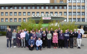
 The group of participants of the 240th ENMC workshop on “The involvement of skeletal muscle stem cells in the pathology of muscular dystrophies” taking place in Hoofddorp from 25-27 January 2019

We acknowledge MD-UK for their co-sponsorship of this workshop.

A full report is published in Neuromuscular Disorders (pdf)